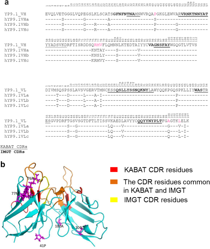
Figure 3. Humanization of the YP9.1 antibody.
(a) Alignment of Fv sequence of YP9.1 and the three versions of humanized YP9.1. The numbers reflect the KABAT system. (b) The structure model of YP9.1 generated by Rosetta (provided by ROSIE Server). The KABAT CDR residues are shown in red; the IMGT residues are shown in yellow. The CDR residues common in KABAT and IMGT are shown in orange. The 41P and 76RMV in VH, 100A and 104L in VL are shown in magenta.

