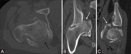
Figure 2 (A-C).

Computed tomography scan of the right hemipelvis. Axial (A), coronal (B), and sagittal (C) images demonstrate a comminuted fracture with a transverse (white arrow) and posterior column (black arrow) pattern of injury involving the right acetabulum
