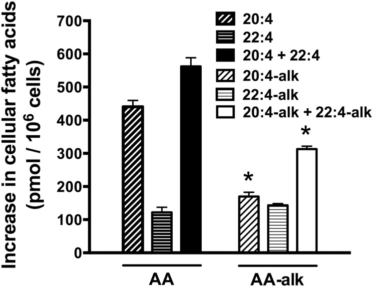
Fig. 3.
The incorporation and elongation of exogenous AA and AA-alk into Jurkat cells. Jurkat cells were incubated with 20 μM AA, 20 μM AA-alk, or their diluent controls for 2 h. Cells were then washed, cellular lipids were extracted, FAs were hydrolyzed and transmethylated, and FAMEs were measured by GC/FID. The results show the increase in the cellular content of AA, 22:4, AA-alk, and 22:4-alk compared with controls following the 2 h incubation period. Data are the means ± SEM of three independent experiments (n = 3). * P < 0.05, different from cells incubated with AA as determined by two-sided Student’s t-tests.

