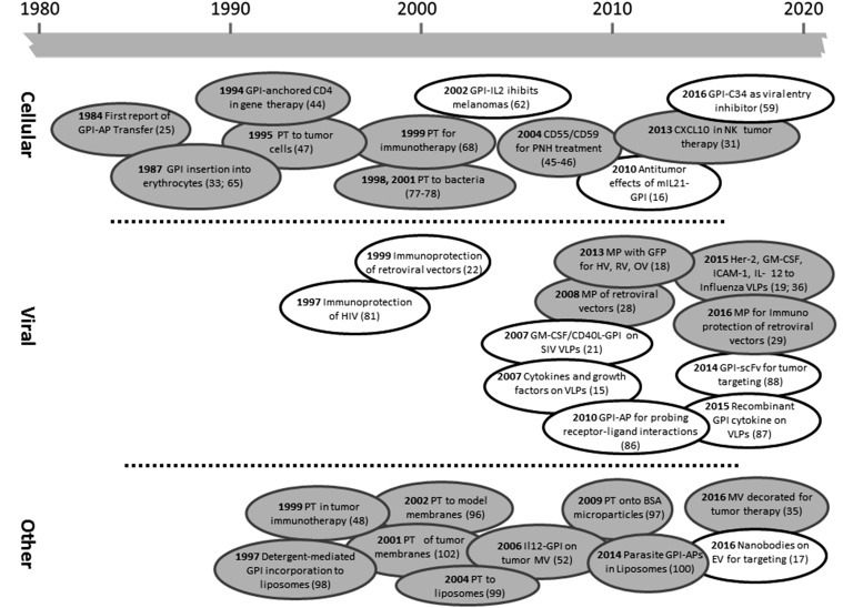
Fig. 3.
Landmarks in GPI-AP membrane engineering. The timeline depicts a selection of key developments in GPI-AP engineering of cellular (top), viral (middle), and other (bottom) lipid bilayer membranes facilitated by GE (clear bubbles) or PE (gray bubbles). EV, extracellular vesicles; HV, herpesviridae; MV, membrane vesicles; OV, orthomyxoviridae; PT, protein transfer; RV, retroviridae; scFv, single chain variable fragment. References to publications can be found in parentheses. For additional information on the proteins used, see Table 1.

