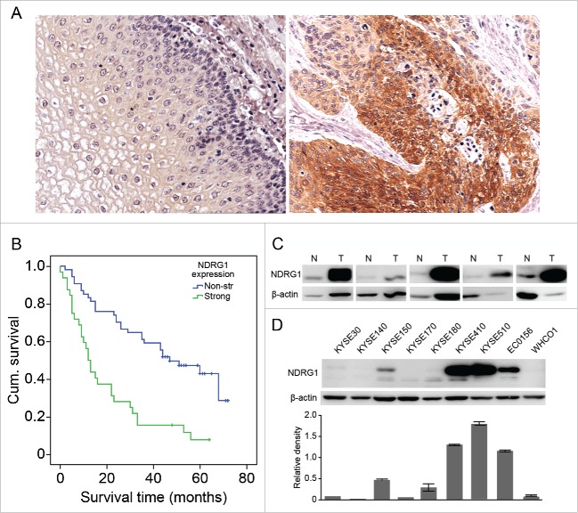
Figure 1.
NDRG1 overexpression was correlated with poor overall survival in esophageal cancer. (A) Expression of NDRG1 was analyzed by immunohistochemical analysis in TMAs containing 78 ESCC tumor and adjacent normal epithelial tissues, with duplicate cores used for each case. The majority of tumor areas strongly express NDRG1 in the cytoplasm. Magnification, 200×. (B) Kaplan-Meier curve combined with Log-rank analysis for patients with ESCC showing weak and strong NDRG1 expression. (C) NDRG1 expression in a subset of ESCC (T) and matched non-neoplastic surgical tissues (N) was analyzed by Western blot analysis. (D) Whole cell protein extracts from 9 esophageal cancer cell lines were subjected to protein gel blot analysis using antibodies against NDRG1. Quantitative values of relative NDRG1 levels were normalized to β-actin (mean ± SD).

