Figure 2.
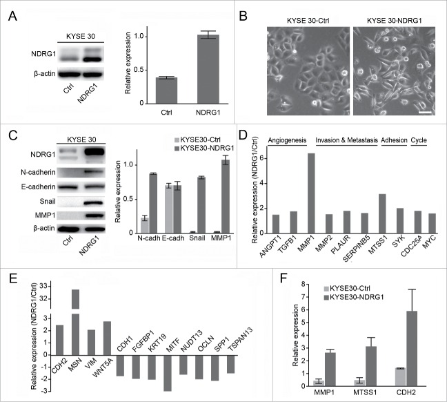
NDRG1 overexpression induces the epithelial mesenchymal transition in esophageal cancer cells. (A) The effect of NDRG1 exogenous expression was analyzed by western blot analysis. Quantitative values of relative NDRG1 levels were normalized to β-actin (mean ± SD). (B) The morphological changes of esophageal cancer cells overexpressed NDRG1. Image at 200x, 20 µm. (C) Whole cell protein extracts from KYSE 30-NDRG1 and KYSE 30-Ctrl cells were subjected to protein gel blot analysis using antibodies against NDRG1, E-cadherin, N-cadherin, Snail and MMP1. Quantitative values of relative protein levels were normalized to actin (mean ± SD). ((D)& E), Relative expression levels of genes associated with angiogenesis, metastasis, adhesion, the cell cycle and the Wnt pathway in KYSE 30 cells overexpressed NDRG1 (KYSE 30-NDRG1) compared with those of mock cells (KYS E30-Ctrl); results are based on analysis in real-time PCR arrays. (F) The mRNA levels of MMP1, MTSS1 and CDH2 were validated in both KYSE 30-NDRG1 and KYSE 30-Ctrl cells by RT-PCR. KYSE 30-NDRG1, NDRG1 overexpression KYSE 30 cells; KYSE 30-Ctrl, KYSE 30 cells transfected with vector control.

