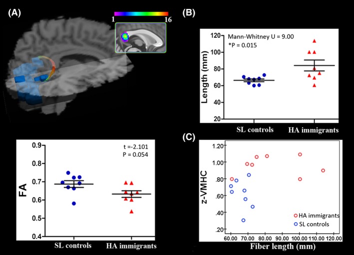
Figure 2.

Diffusion tractographic images from a single control subject show between‐group comparison for commissural fiber parameters. (A) Diffusion tractographic images from a single control subject. Fibers connecting the bilateral visual cortex are located in the splenium of the corpus callosum. The inset shows the probabilistic maps of the commissural tract constructed with data from 16 subjects. (B) Scatterplots show the between‐group comparison for the commissural fiber parameters of fractional anisotropy (FA) and fiber length. *P < 0.05, Bonferroni corrected. (C) A significant correlation between mean z‐VMHC index within homotopic visual areas and the length of their commissural fibers. Spearman rho = 0.565, P = 0.023. A longer path length of the fibers connecting the bilateral visual cortex corresponds to stronger interhemispheric functional synchronization. SL, sea level; HA, high‐altitude.
