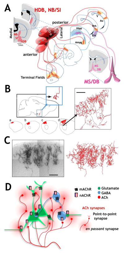
Figure 1. Functionally modular projection patterns, exotic axonal morphologies and diverse ACh release-receptor interactions contribute to complex spatio-temporal dynamics of ACh signaling by basal forebrain cholinergic neurons.
(see text and Reviews by Munoz & Rudy, 2014; Picciotto et al., 2012; Sarter, 2016; Zaborzsky et al., 2015).
A. Schematic of projection patterns of basal forebrain cholinergic neurons. Left hand side: schematic of coronal sections indicating the approximate anterior to posterior and medial to lateral distribution of the HDB (horizontal limb of the diagonal band) and NB/SI (Nucleus Basalis/Substantia innominata). Anterior medial BFCNs within these nuclear groups project to medial frontal cortical targets whereas posterior located cholinergic neurons project to more posterior targets such as the BLA and perirhinal cortex. Right hand side: Medial septal (MS) and vertical limb of the diagonal band (VDB) neurons provide cholinergic input to the hippocampus and entorhinal cortex.
B. and C. Axonal morphology of fully reconstructed basal forebrain cholinergic neurons and the extensive terminal arborization formed in cortex. Adapted with permission from Wu et al., 2014 https://creativecommons.org/licenses/by/3.0/.
D. Schematic representation of both point-to-point (focused, triangular) and en passant (broad circular) mechanisms by which ACh is released in the CNS, thereby effecting both glutamatergic (green) and GABAergic (blue) neuronal excitability. Such release profiles may correspond to the more rapid and transient responses and the slower, longer lasting modulatory effects of ACh, respectively (see text for discussion). Also shown are representative distributions of both muscarinic (depicted as 7 TM squiggles) and nicotinic (represented as single tubes) AChRs at pre, post and peri synaptic sites. Both mAChR and nAChR subtypes at each of these locations contribute to the direct and indirect mechanisms by which ACh can alter synaptic excitability (see text for discussion).

