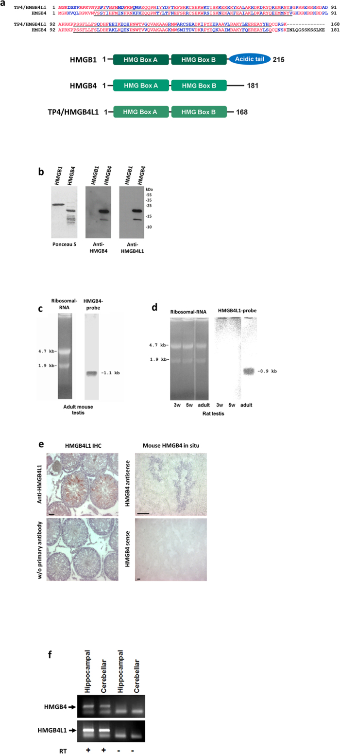
Figure 1. Characterization of HMGB4 and HMGB4L1.

(a) Alignment of rat TP4/HMGB4L1 and HMGB4 amino acid sequences. HMGB-boxes A and B are underlined. Red letters indicate identical amino acids. An alternative allele in position 34 of TP4/HMGB4L1 is either a tyrosine or a isoleucine. Amino acids marked in italics have been identified by amino terminal amino acid sequencing7. Domain structures and number of amino acids of rat HMGB1, HMGB4 and TP4/HMGB4L1 are shown in the schematic picture. (b) Western –blot of recombinant mouse HMGB4. Recombinant HMGB4 was detected with Ponceau S –staining and with anti-HMGB4 and anti-HMGB4L1 antibodies. The antibodies did not detect recombinant HMGB1. (c) Northern Blot -analysis of the mouse HMGB4 transcript. Total RNA samples were isolated from adult mouse testes and analyzed via Northern Blot, using a probe derived from the entire coding sequence of mouse HMGB4. The probe detected a 1.1 kb band. Ethidum bromide stained ribosomal RNA is shown. (d) Northern Blot -analysis of rat HMGB4L1 transcript. Total RNA samples were isolated from adult rat testes and from developing rat testes, and analyzed via Northern Blot, using a probe derived from the entire coding sequence of rat HMGB4L1. The probe detected a 0.9 kb band in samples derived from testes of sexually mature rat. Ethidum bromide stained ribosomal RNA is shown. 3 w = 3 week old rat, 5 w = 5 week old rat. (e) Immunohistochemical staining of rat HMGB4L1 protein and in situ hybridization of mouse HMGB4 mRNA in adult testes. Anti-HMGB4L1 polyclonal antibody staining revealed intense HMGB4L1 expression in elongated spermatids (red). Haematoxylin was used as a counterstain. Control sections were stained without the primary antibody. HMGB4 mRNA localized to round and elongated spermatids. The control section shows adult mouse testes incubated with the sense probe. Bars represent 50 μm. (f) Expression of HMGB4 and HMGB4L1 mRNA in cultured rat neurons. Neuronal cells from the hippocampus and the cerebellum were cultured, and both HMGB4 and HMGB4L1 expression (arrows) was detected with RT-PCR. +RT = reverse transcribed, -RT = without reverse transcription.
