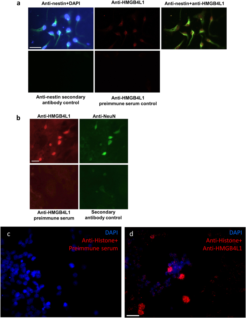
Figure 6. Expression of HMGB4 and HMGB4L1 in cultured primary cells of rat brain.
(a) Immunofluorescence staining of nestin and HMGB4L1 in 1d differentiated rat neuronal cells in vitro. Neurospheres were allowed to adhere and differentiate for 1 day and cells were immunofluorescently stained with anti-nestin and anti-HMGB4L1 antibodies. Cell nuclei were stained with DAPI. The staining controls for anti-nestin or anti-HMGB4L1 staining were done without primary antibody or with preimmune serum, respectively. Scale bar = 30 μm. (b) Immunofluorescence staining of HMGB4L1 and NeuN in 14d differentiated rat neuronal cells in vitro. Neurospheres were allowed to differentiate for 14 days and cells were immunofluorescently stained with anti-NeuN and anti-HMGB4L1 antibodies. Staining controls for anti-HMGB4L1 and anti-NeuN were done with pre-immune serum or without primary antibody, respectively. Scale bar = 20 μm. (c,d) Proximity ligation assay of neurospheres with anti-PAN-Histone and anti-HMGB4L1 antibodies. Cell nuclei of cultured neurospheres were stained with DAPI and a proximity ligation assay was performed with anti-PAN-Histone antibodies and preimmune control serum (c) or with anti-PAN-Histone antibodies and anti-HMGB4L1 antibodies (d). Scale bar = 20 μm.

