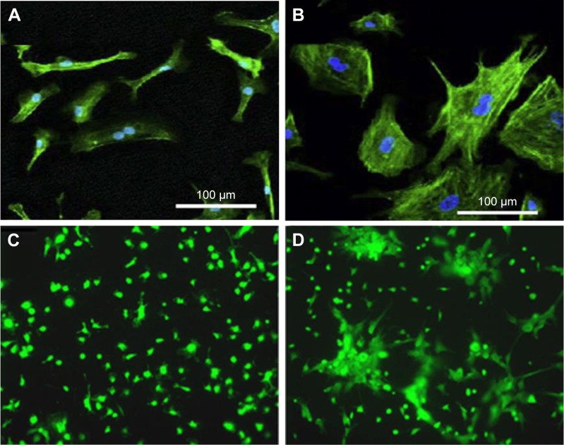
Figure 2.
F-actin and nuclear stains of BAEC.
Notes: F-actin (green) and nuclear (blue) stains of BAEC grown on (A) TNTs versus (B) flat surfaces for 24 hours. Fluorescence microscopy images (×10) of live marrow stromal cells stained with calcein on (C) Ti and (D) TNT surfaces. (A and B) Reprinted from Biomaterials, 30, Peng L, Eltgroth ML, LaTempa TJ, Grimes CA, Desai TA, The effect of TiO2 nanotubes on endothelial function and smooth muscle proliferation, 1268–1272,55 Copyright (2009), with permission from Elsevier. (C and D) Reprinted from Biomaterials, 28, Popat KC, Leoni L, Grimes CA, Desai TA, Influence of engineered titania nanotubular surfaces on bone cells, 3188–3197,57 Copyright (2007), with permission from Elsevier.
Abbreviations: BAEC, bovine aortic endothelial cell; TNT, titania nanotube.

