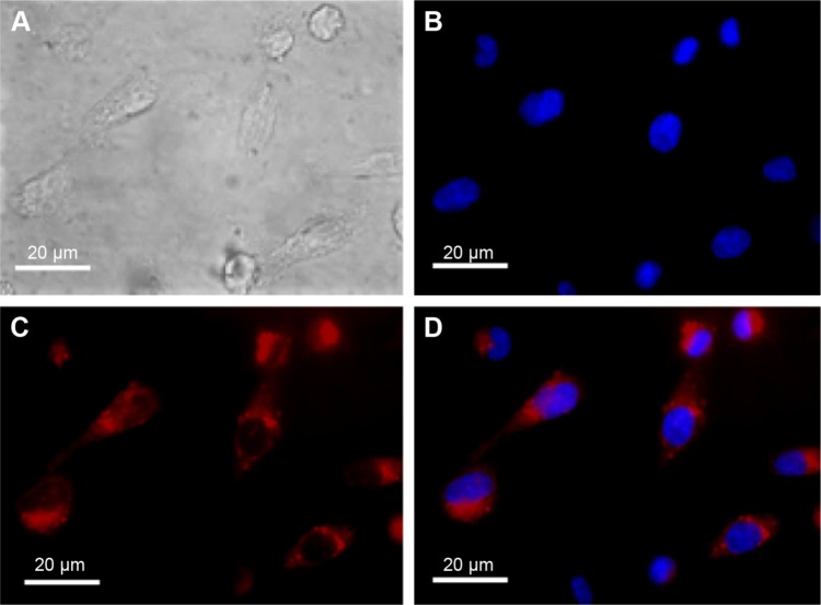
Figure 3.
Fluorescence cell imaging.
Notes: MDA-MB-231 cells were incubated with NIR-PluS NPs (NP-7; 100 nM) at 37°C for 24 hours, to allow NIR-PluS NP cell internalization before microscopy analysis. Representative cell images are shown. DAPI staining was carried out to visualize cell nuclei, which appear in blue, while NIR-PluS NPs appear in red. (A) Bright field image; (B) DAPI nuclear staining; (C) fluorescence emission; and (D) overlaid images (bar =20 µm).
Abbreviations: NIR, near infrared; NIR-PluS NPs, NIR-emitting pluronic-silica nanoparticles.

