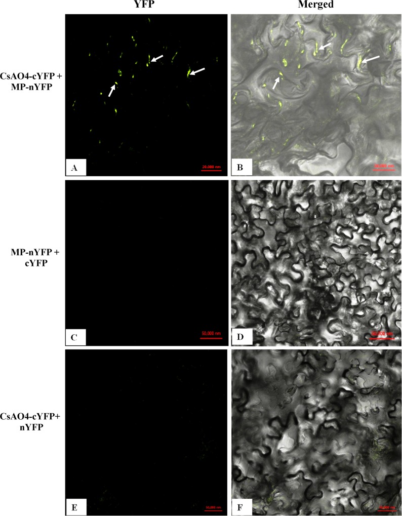
Fig 3. In planta interaction between CsAO4 and CMV MP by BiFC.
The lower surface of N. benthamiana leaves was observed under the confocal microscope for fluorescence from YFP: A) and B) CsAO4-cYFP and MP-nYFP. C) and D) MP-nYFP and cYFP. E) and F) CsAO4-cYFP and nYFP. YFP reconstitution observed in A and B showed punctate sites around the cell wall periphery. Confocal images were merged with bright field images. Fluorescence was detected 48 h post agroinfiltration. Scale bars are shown in the figures.

