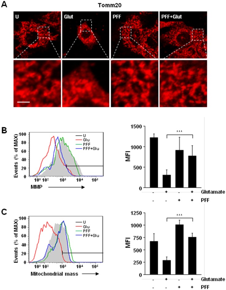
Fig 5. Glutamate-induced mitochondrial dysfunction in PC12 cells was improved by PFF treatment.
PC12 cells were stimulated with glutamate (5 mM, 24 h) in the presence or absence of PFF (10 μM). (A) Cells were immunolabelled with a Tomm20 antibody, followed by the addition of Cy3-conjugated secondary antibody. Representative immunofluorescence images (scale bars = 10 μm). (B) Mitochondrial membrane potential (ΔΨm) was measured under the indicated conditions using the ΔΨm-sensitive fluorochrome MitoTracker Red CMXRos and flow cytometric analysis. (C) MitoTracker fluorescence signals for mitochondrial mass were measured using flow cytometric analysis. (Left in B and C) Representative histogram from three independent replicates. (Right in B and C) Bar graph shows ΔΨm (B) or mitochondrial mass (C) mean fluorescence intensities. Data represent the means and SD of three independent experiments. ***p < 0.001 vs. the control group. U, untreated condition. Glut, glutamate.

