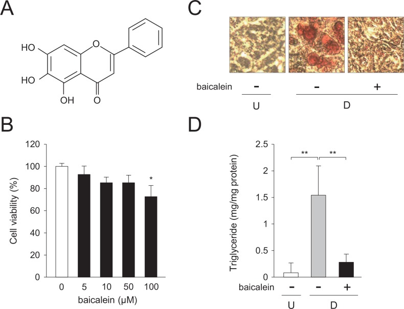
Fig 1. Decrease in intracellular lipid accumulation in 3T3-L1 cells by baicalein.
A, Structure of baicalein. B, Cell toxicity of baicalein toward 3T3-L1 cells. Cells were cultured for 6 days in DMEM containing various concentrations of baicalein (0–100 μM). Data are the means ± S.D. *p<0.05, as compared with the vehicle (0 μM). C, Oil Red O staining of baicalein-treated 3T3-L1 cells. Cells (undifferentiated cells: U) were differentiated into adipocytes (differentiated cells: D) for 6 days in DMEM without or with baicalein (0 or 50 μM). Intracellular lipid droplets were stained with Oil Red O. D, Baicalein-mediated suppression of the intracellular triglyceride level of 3T3-L1 cells. Cells (undifferentiated cells: U; white column) were differentiated into adipocytes (differentiated cells: D) for 6 days in DMEM without (gray column) or without baicalein (50 μM; black column). Data are presented as the means ± S.D. **p<0.01, as indicated by the brackets.

