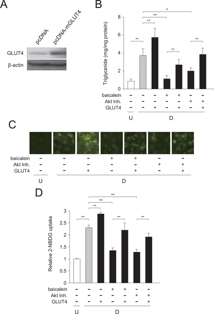
Fig 9. Expression of GLUT4 in baicalein- or Akt inhibitor-treated 3T3-L1 cells.
A, Expression of the recombinant mouse GLUT4 protein in 3T3-L1 cells. 3T3-L1 cells were transfected with GLUT4 (pcDNA-mGLUT4) or the empty (pcDNA) vector. GLUT4 levels were detected by Western blot analysis using cell extracts (15 μg/lane). B, Intracellular triglyceride level in GLUT4-transfected cells. 3T3-L1 cells were transfected with GLUT4 vector by electroporation. After 24 h, cells (undifferentiated cells: U; white column) were differentiated into adipocytes (differentiated cells: D; gray column) for 6 days and treated with baicalein or an Akt inhibitor (Akt Inh.; black columns) for the initial 1.5 h of adipogenesis. Data are presented as the means ± S.D. from three experiments. *p<0.05, **p<0.01, as indicated by the brackets. C, Change in glucose uptake in GLUT4-transfected cells. 3T3-L1 cells were cultured as described in the legend of Fig 9B. Cells were then incubated with fluorescent 2-NBDG, and then observed under a fluorescence microscope. The results are representative from three experiments. D, Measurement of glucose uptake in GLUT4-transfected cells. Data are presented as the value relative to that of the undifferentiated cells and are shown as the means ± S.D. from three experiments **p<0.01, as indicated by the brackets.

