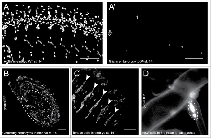
Figure 1.

A-A′) Confocal projections of wild type (A) and gcm LOF (A′) embryos at stage 14 immunolabelled with the Repo glial marker, anterior to the left, latero-ventral views. Note the absence of glia in the gcm LOF embryo. B, C) Confocal sections of a gcm > GFP transgenic embryo at stage 14 labeled with the anti-GFP antibody, anterior to the left, dorso-lateral views. The section was acquired in the inner layers of the embryo to reveal the circulating hemocytes, outlined by a dashed line in (B) and in the outer layers to reveal the tendon cells indicated by arrowheads in (C). The scale bar in (A–C) represents 50 µm. D) Tracheal branch of a gcm > GFP 3rd instar larva analyzed by epifluorescence microscopy (200x magnification). The INKA cells are recognizable by their position and are outlined by the dashed line.
