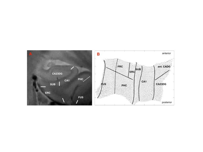
Figure 1.
Cortical unfolding. After oblique coronal MRI scanning and manual segmentation of white matter and CSF on the T2 weighted MRI sequence, the resulting gray matter volume is computationally unfolded and flattened based on metric multidimensional scaling [right hemispheric flatmap shown, B]. Boundaries between the subregions are delineated on the original high-resolution MRI sequence (A) and later mathematically projected to flat map space. CA23DG=cornu ammonis fields 2,3 and dentate gyrus (the anterior part of the cornu ammonis fields and dentate gyrus [ant. CADG] is part of the CA23DG region), CA1=CA field 1, SUB=subiculum, ERC=entorhinal cortex, PRC=perirhinal cortex, PHC=parahippocampal cortex, FUS=fusiform cortex (fusiform boundary depicts the medial fusiform vertex).

