Figure 5. Long-time retrieval of remote fear memory reactivated the hippocampus (HP).
(A) Representative immunohistochemical staining of the anterior cingulate cortex (ACC) and CA1 c-fos-positive cells from the indicated group. Scale bars = 100 μm. Two groups were trained with a foot shock (trained group) and the remaining two groups did not receive a foot shock (untrained group). Twenty-four hr (recent memory) or four weeks (remote memory) after the training, mice were placed back in the training context for 3 or 10 min for short or long memory recall, respectively (re-exposure). (B) The c-fos expression in the ACC (layers II/III, and V) and the hippocampus [CA1, CA3, and the dentate gyrus (DG) regions]. N = 10 for each group. The c-fos expression in the trained groups was expressed as a percentage relative to the untrained groups. (C) Ten min re-exposure to the context four weeks after training [vehicle-treated group (VEH), n = 11, lidocaine-treated group (LIDO), n = 11]. Error bars, SEM. *p<0.05. The results of the statistical analyses are presented in Figure 5—source data 1.
DOI: http://dx.doi.org/10.7554/eLife.17464.011

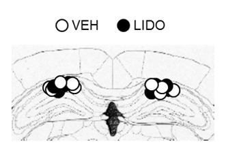
Figure 5—figure supplement 1. The cannula tip placement in the hippocampus.