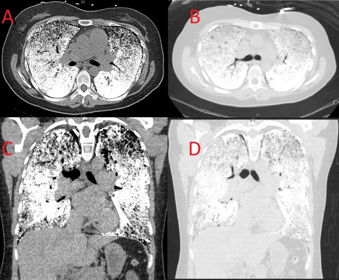
Figure 2. Computed tomography of the chest.
A: Axial view, intense calcification of the interstitium and pleural serosa. B: Axial view lung window, intense calcification giving a “white lung” appearance. C: Coronal view, intense calcification of the interstitium and pleural serosa sparing the lung apices. D: Coronal view lung window, dense calcification of the lower lung fields with moderate calcification of the apices.

