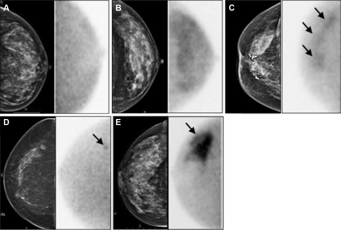
Fig. 2.
Examples of MBI according to BI-RADS classification [12] displayed together with corresponding mammography. Left craniocaudal view (a) showing homogeneous uptake (BI-RADS I); left craniocaudal view (b) showing diffusely increased uptake (BI-RADS II); right craniocaudal view (c) showing multiple patchy areas of uptake (BI-RADS III) pointed by arrows; right craniocaudal view (d) showing small focal area of increased uptake (BI-RADS IV, arrow); right craniocaudal (e) showing intense uptake (BI-RADS V, arrow)

