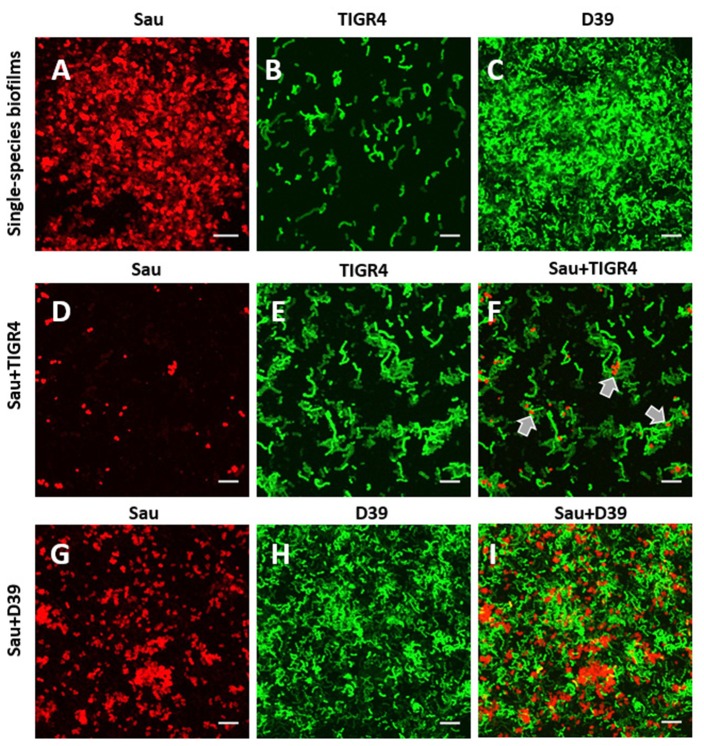
Figure 6.
Confocal studies of Sau coincubated with Spn strains. (A) Sau, (B) SPJV09 (TIGR4), or (C) SPJV01 (D39), or mixtures of Sau and SPJV09 (D–F) or Sau and SPJV01 (G–I) was inoculated into an eight-well slide and incubated for 4 h at 37°C. Biofilms were fixed with 2% PFA and stained with an anti-Sau antibody followed by an Alexa 555-labeled anti-rabbit secondary antibody (red). Spn strains were expressing the green fluorescent protein. Preparations were analyzed by confocal microscopy. A representative xy optical section is shown. Bar = 20 μm. Gray arrows point out areas where Sau and TIGR4 are located.

