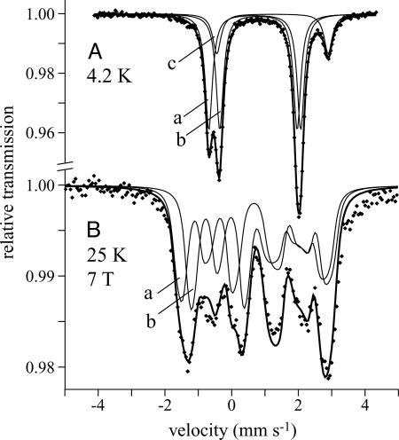
Fig. 5.
Mössbauer spectra of the [2Fe-2S]0-H+ Rieske cluster (pH 7). (A) B = 0, 4.2 K. (B) B = 7 T perpendicular to the γ-beam, 25 K. A was modeled by using Lorentzian doublets (see Table 1), and the lines shown in B are spin-Hamiltonian simulations (see text). The “nested” configuration of doublets is shown, and subspectra a and b represent the cysteine- and histidine-coordinated centers, respectively. Subspectrum c represents contamination from cluster degradation (16%).

