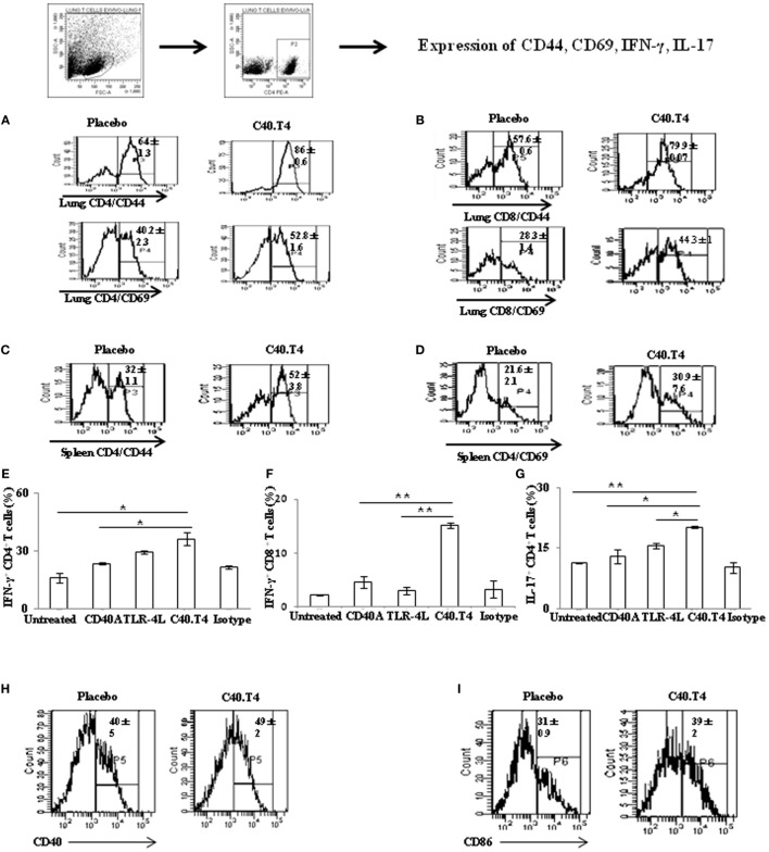
Figure 7.
C40.T4 treatment bolstered the T cells immunity in mice exposed to Mtb. Mtb-infected animals were administered C40.T4. After 50 days, mice were sacrificed and lymphocytes were isolated and cultured with purified protein derivative (6 μg/ml) for 48 h. Later, cells were stained for the expression of (A) CD44/CD69 on CD4 T cells isolated from the lungs; (B) CD44/CD69 on CD8 T cells isolated from the lungs; (C,D) CD44/CD69 on CD4 T cells isolated from the spleens. Number in the histograms indicates the percentage of CD44hi and CD69hi on CD4 and CD8 gated population. Splenocytes stimulated in vitro with PPD were stained for expression of (E) IFN-γ on CD4 T cells; (F) IFN-γ on CD8 T cells; (G) IL-17 on CD4 T cells. The bar diagrams illustrate percent population and results expressed as mean ± SD. DCs isolated from the lungs of Mtb-infected animals treated with C40.T4 were stained for the expression of (H) CD40; (I) CD86 and on CD11c + DCs. Number in the histogram indicates the mean ± SD of CD11c+ DCs expressing CD40 and CD86 population. The data are representative of two independent experiments (n = 4 mice/group). “*,” “**,” and “***” indicate p < 0.05, p < 0.01, and p < 0.001, respectively.

