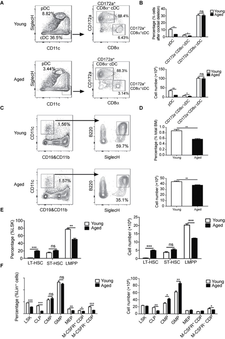
Figure 1.
PDC and CD172a−CD8α+cDC were decreased in aged mice. (A) DC from the spleens of young and aged mice were enriched, and the DC subsets were analyzed by flow cytometry. The plots show the percentages of DC subsets in the spleens of young and aged mice. (B) The percentages and cell numbers of DC subsets in the spleens of young and aged mice. (C) The plots show the percentages of pDC in the BM of young and aged mice. (D) The percentages and cell numbers of pDC in the BM of young and aged mice. (E) Lin− cells enriched from the BM of young and aged mice were analyzed by flow cytometry. The graphs show the percentages and cell numbers of HSC populations in young and aged mice. (F) The percentages and cell numbers of LSK, CMP, CLP, GMP, MEP, M-CSFR+ CDP, and M-CSFR− CDP in young and aged mice. Data are representative of three independent experiments. Error bars represent SEM (*P < 0.05, **P < 0.01, ***P < 0.001).

