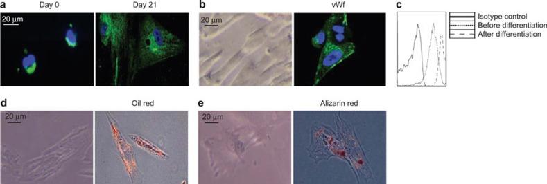
Figure 2.
Ex vivo differentiation of CD34loCD133lo cells. CD34loCD133lo cells were purified and cultured in various differentiation media. (A) CD34loCD133lo cells were cultured in hepatocyte differentiation medium. Representative images of cultured CD34loCD133lo cells immunofluorescently stained on day 0 and 21 post-culture for human ALB (green) and DAPI (blue). (B) Representative phase-contrast micrographs (left) and immunofluorescence stains for human Von Willebrand Factor (vWf) (green) and DAPI (blue) (right) are shown for endothelial cell differentiation cultures after 7 days in vitro. (C) Cells from endothelial cell differentiation cultures were stained with a CD31 antibody and analyzed by flow cytometry. The histograms of the CD31 stains of CD34loCD133lo cells before and after differentiation are shown. (D) Representative phase-contrast micrographs (left) and stains for Oil Red O (red) (right) are shown for adipocyte differentiation cultures after 7 days in vitro. (E) Representative phase-contrast micrographs (left) and stains for Alizarin Red S (red) (right) are shown for osteocyte differentiation cultures after14 days in vitro. The scale bar applies to all panels.

