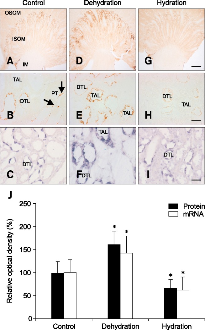
Fig. 2. Light micrographs illustrating OPN immunostaining and in situ hybridization in the kidney from control (A–C), dehydrated (D–F) and hydrated (G–I) rats. The OPN immunostaining and hybridization signal increased remarkably in dehydrated animals, while it decreased in hydrated animals relative to control animals. OPN expression was primarily observed in the tubular profiles in the inner stripe of the outer medulla (ISOM). Note that the OPN immunostaining and hybridization signal were not only observed in the descending thin limb of Henle's loop (DTL), but also in the thick ascending limb (TAL) in dehydrated animals (E and F). Arrows indicate an abrupt transition from the OPN-negative proximal tubule (PT) to the OPN-positive descending thin limb of Henle's loop. Quantitative analysis of OPN protein and mRNA expression (J). OSOM, outer stripe of the outer medulla; IM, inner medulla. *p < 0.05 by two-tailed t-test versus control. Scale bars = 1 mm (G), 20 µm (H and I).

