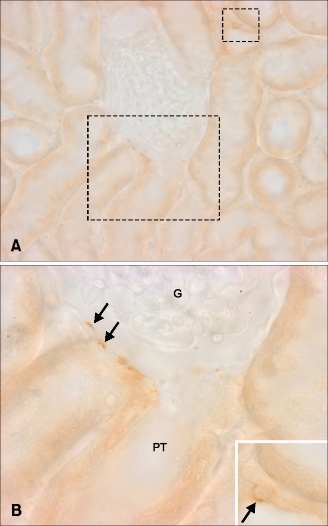
Fig. 3. Light micrographs illustrating OPN immunostaining in the cortex (A). Occasionally, OPN expression was observed in Bowman's capsules (B, arrows) and distal tubules (inset, arrow). Rectangles mark areas magnified. G, glomerulus; PT, proximal tubule.

