
Figure 5.
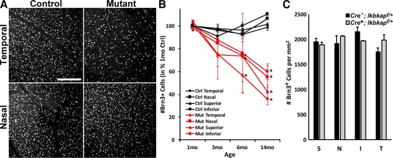
Loss of Ikbkap in RGCs caused slow, progressive RGC degeneration. A, Representative Brn3 (RGC marker) staining in the temporal and nasal retinas at 6 months. Images were taken at 1 mm from ONH. B, The number of Brn3+ RGCs in Ikbkap CKO (mutant) retinas was counted. Significant loss of Brn3+ cells was observed in temporal and superior retinas at 6 months, which progressively spread into entire retinas by 14 months. C, The numbers of Brn3+ RGCs were counted in each quadrant of 6- to 8-month-old Cre-;Ikbkapf/+ and Cre+;Ikbkapf/+ retinas at 1 mm from the ONH. There was no significant decrease in the number of RGCs in Cre+;Ikbkapf/+ compared to Cre-;Ikbkapf/+retinas, demonstrating that Cre expression itself and/or loss of one Ikbkap allele did not cause RGC degeneration. S, superior; N, nasal; I, inferior; T, temporal. Error bars represent SEM (n = ≥4 per point for B; n = 3 for C). *p < 0.05 with t-test. Scale bars, 100 μm (A).