Figure 6.
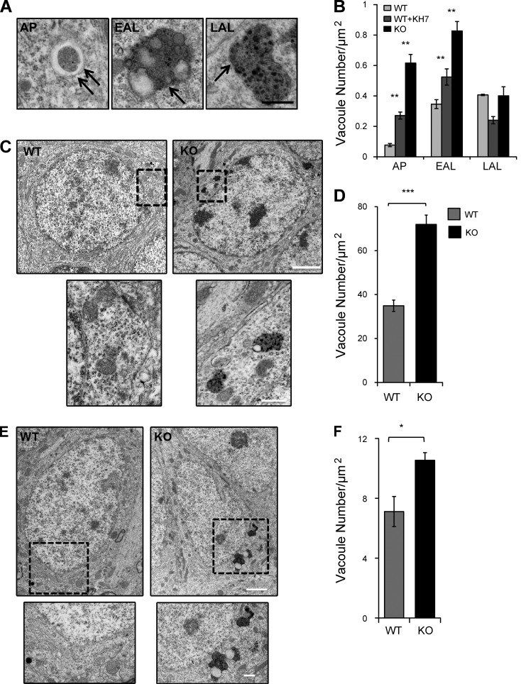
Electron-dense AVs accumulate in the absence of sAC activity. (A) AVs were subcategorized based on their morphology. AP: double membrane structures and/or double membrane structure containing undigested organelles; EAL: single membrane structures containing partially digested electron-dense material; LAL: single membrane structures containing amorphous electron-dense material. Double arrows represent double membrane, and single arrow represents single membrane. (B) Quantitative analysis of the number of AVs in WT, WT+KH7, and sAC KO MEFs. n = 10 cells/condition, two independent experiments. **, P < 0.02. (C) Representative electron micrographs of hippocampal dentate granule cells in WT and sAC KO aged mice. High-magnification views of the boxed areas below show examples of AVs in KO mice. (D) Quantitative analysis of the number of AVs in WT and sAC KO granular cells of the dentate gyrus. n = 3 mice/condition; n = 10 cells/mouse. ***, P < 0.002. (E) Representative electron micrographs of the pyramidal cells of the CA1 in WT and sAC KO aged mice. High-magnification views of the boxed areas below show examples of AVs in KO mice. Bars: (A, C [bottom], and E [bottom]) 500 nm; (C and E, top) 2 µm. (F) Quantitative analysis of the number of vacuoles in WT and sAC KO pyramidal cells of the CA1 of the hippocampus. n = 3 mice/condition; n = 10 cells/mouse. *, P < 0.05. All values are given as mean ± SEM.

