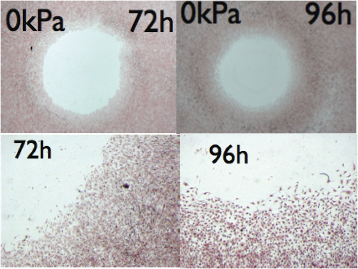
Figure 1. Human melanocytic cells (primary tumour) were plated on 33 cm2 Petri dish with at the center a disk (32 mm2) at confluence.
The day after, the disk was removed and cells were allowed to keep migrating and dividing, before they were fixed with 3.7% paraformaldeide and stained with hematoxillin/eosin solution, at different times. On top, typical experiment, at two different times, on bottom, details from the same images with X5 magnification, compared to the top (Leica MZFLIII mounted with a camera Leica DFC320). Observe the diffuse front and an increase of noise after 4 days. From publication12 with permission.

