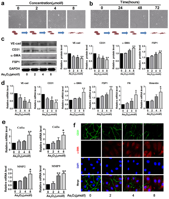
Figure 4. As2O3 triggers EndMT in human aortic endothelial cells (HAECs).
(a) Morphological changes of endothelial cells exposed to different concentrations of As2O3. (b) Morphological changes of endothelial cells treated with As2O3 (2 μmol/l) for 0, 24, 48 or 72 hours. (c) Western blotting results for relative protein levels of VE-cad, CD31, α-SMA and FSP1 in As2O3-treated HAECs. GAPDH was used as an internal control. (d) Relative expression levels of endothelial markers (VE-cad, CD31) and mesenchymal markers (α-SMA, FSP1, FN and Vimentin) were compared by qRT-PCR between control groups and As2O3-treated groups. FN, fibronectin. (e) Relative mRNA levels of fibrosis-related genes Col1a, Col3a, mmp2 and mmp9. (f) Representative confocal microscopy images showing staining of endothelial marker CD31 and mesenchymal marker α-SMA in As2O3-treated HAECs. Scale bar = 30 μm. *p < 0.05, **p < 0.01 vs. untreated condition (0 μmol/l As2O3). No significant difference was observed between the different concentrations groups. Data are represented as mean ± SEM, n = 3–5.

