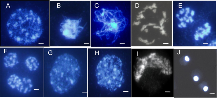
Figure 4. Male meiosis in fertile and sterile pollen mother cells (PMCs) prepared using the spreading technique and stained with DAPI.
(A–F), meiosis in fertile PMCs; (G–J), meiosis in sterile PMCs; (A,G), leptotene. several bright stained chromatin centers were visible; (B,H), zygotene; (C) wild-type fertile pachytene showing full chromosome synapsis; (D) fertile diakinesis showing moderately condensed, unaligned bivalents; (E) telophase I, dyads were formed; (F) telophase II, tetrads were formed; (I) arrangement of chromosomes was disordered, and chromosomes formed the crescent-like structure; (J) the final arrested PMCs. Chromosomes remain together as a diffused mass. Neither meiosis I nor meiosis II is completed in these MMCs, showing meiosis being completely arrested. Scale bars = 5 μm.

