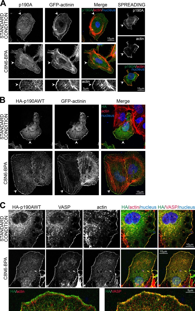
Figure 1.
p190A localizes to membrane protrusions in Huh7 cells. (A) As a standard condition, Huh7 cells were plated on glass coverslips and then fixed after overnight culture. Lamellipodium growth was either stimulated by spreading on fibronectin for 30 min (top) or by treatment with 100 µM C8N6-BPA (middle). Huh7 cells were transfected with α-actinin–GFP used as a lamellipodium marker, fixed, and stained for p190A (green), F-actin (red), and nuclei (blue). Bottom panel shows enlargement of C8N6-BPA condition. (B) Huh7 cells were transfected with HA-p190AWT and α-actinin–GFP, fixed, and stained for HA tag (green), F-actin (red), and nuclei (blue). (C) Huh7 cells were transfected with HA-p190AWT, fixed, and stained for HA tag (green), VASP (red) or F-actin (red), and nuclei (blue). Bottom panel shows enlargement of C8N6-BPA condition. (A and B) Arrowheads show membrane ruffles with p190A/HA-p190AWT localization.

