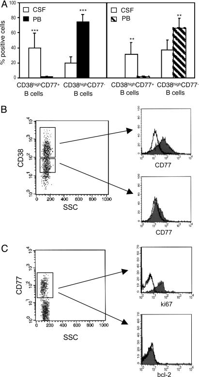
Fig. 3.
Identification of CD19+, CD38high, CD77+, Ki67+, Bcl-2– centroblasts in the CSF of patients with MS or OIND. (A) Cells from CSF and PB of 10 MS (Left) and 10 OIND (Right) patients were stained with CD19 and CD38 mAbs in combination with CD77 and analyzed by flow cytometry setting the gate on CD19+ cells. Coexpression of CD77 and CD38 was determined by setting the marker on CD38high cells, which represent bona fide GC B cells (B). Results are mean percent positive cells ±SD. ***, P = 0.0005; **, P = 0.001. (B) Cells from the CSF of a MS patient were stained with CD19, CD38, and CD77 mAbs and analyzed by gating first on CD19+ (data not shown) and then on CD38high or CD38low cells (Left). CD77+ cells were detected in the CD38high (Upper Right) but not in the CD38low (Lower Right) B cell fraction. A representative experiment is shown. (C) Cells from the CSF of a MS patient were stained for surface CD19 and CD77, as well as intracellular Ki67 or Bcl-2, and analyzed by gating first for CD19 (data not shown) and then for CD77 (Left). CD77+ B cells expressed Ki67 (Upper Right) but not Bcl-2 (Lower Right). A representative experiment is shown.

