Figure 2.
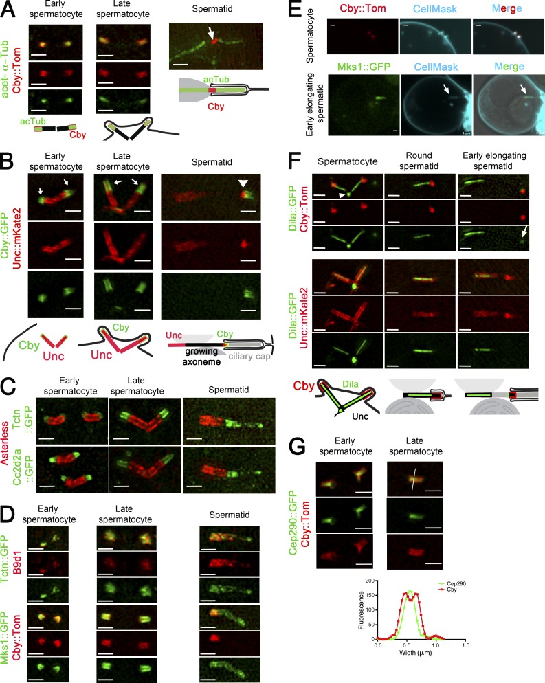
Localization of TZ components in Drosophila male germ cells. (A–D, F, and G) 3D-SIM imaging of male germ cells. Recapitulating schemes are based on Fig. 1. (A) Acetylated tubulin (acTub) and Cby overlap in spermatocytes. In elongating spermatids, Cby is restricted to the ring centriole (arrow), and acetylated tubulin stains the axoneme proximal and distal to the ring centriole. (B) Unc labels the BB, and Cby and Unc colocalize along the TZ/cilia (arrows). The ring centriole labeled with Unc and Cby (arrowhead) separates from the BB in elongating spermatids. (C and D) Tctn, Cc2d2a, B9d1, and Mks1 colocalize at the tip of the centrioles (Asl, red) and overlap with Cby in spermatocytes. In early elongating spermatids, all MKS components cover the entire ciliary cap, whereas Cby is restricted to the ring centriole. (E) Live confocal imaging of Cby and Mks1. Membranes are labeled with CellMask. Cby labels entirely the cilia/TZ in spermatocytes, and Mks1 labels the entire ciliary cap (arrows) in spermatids. (F) Dila covers the entire lumen of centriole and projects into the TZ/cilia marked by Cby or Unc in spermatocytes and round spermatids. Dila is at the base of centrioles (arrowhead), and a small fraction migrates with the ring centriole in the spermatid (arrow; gamma was adjusted to 1.35 for the green layer). (G) Cby surrounds Cep290 both at the TZ/cilia and at the ring centriole as observed on a scan profile of fluorescence intensity (grayscale units) across (white line) the TZ/ring centriole. Bars, 1 µm.

