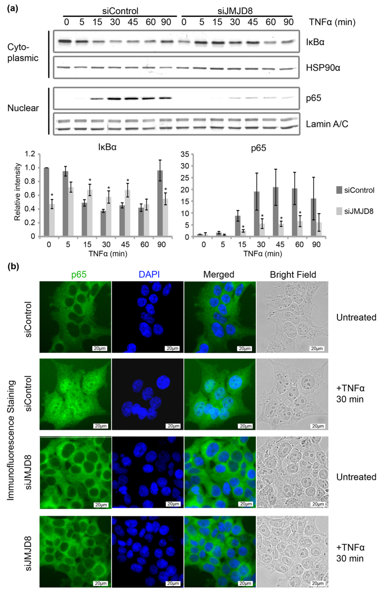
Figure 2. JMJD8 deficiency reduces TNF-induced IκBα degradation and p65 translocation.
(a) Control and JMJD8 knockdown HEK293T cells were treated with 10 ng/ml of TNFα for 0, 5, 15, 30, 45, 60 and 90 minutes. Cytoplasmic and nuclear fractions were prepared and immunoblotted for IκBα and p65. HSP90α and Lamin A/C were used as cytoplasmic and nuclear loading controls respectively. Relative intensity of bands were quantified using the Image Lab (BioRad)/ImageJ software, were normalized to HSP90α or Lamin A/C, and shown in relative to 0 minute of siControl (n = 3). (b) Control and JMJD8 knockdown HEK293T cells were treated with 10 ng/ml of TNFα for 30 minutes. P65 localization was visualized with an immunofluorescence assay. Images were acquired with an Olympus IX71 fluorescence microscope. Scale bar: 20 μm. (n = 3). Data represent means ± SD. (*p > 0.05). Full-length blots are presented in Supplementary Fig. S4.

