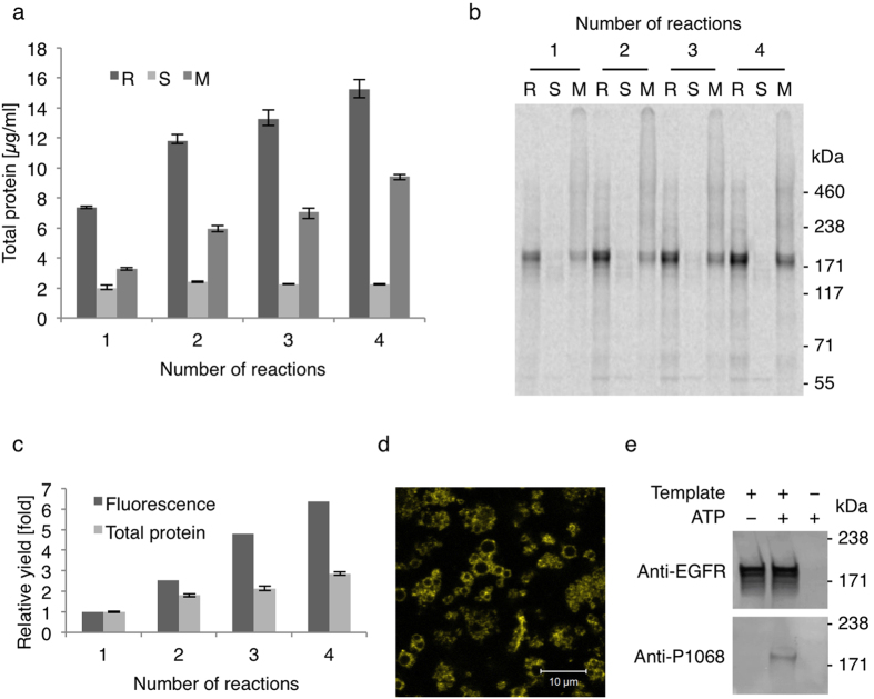
Figure 1. Enrichment of functional EGFR-eYFP in the Sf21 microsomal fraction by repetitive cell-free synthesis.
(a) Total protein yields after each cell-free reaction in the complete reaction mixture (R), the supernatant fraction (S) and the microsomal fraction (M). Error bars represent the standard deviation of triplicate analysis. (b) Autoradiography of corresponding samples after electrophoretic separation under denaturing conditions. (c) Yields of total protein and eYFP fluorescence after each cell-free reaction relative to the first reaction. (d) Confocal fluorescence image of EGFR-eYFP in microsomal fraction taken under hypoosmotic buffer conditions. (e) Western Blot of microsomal fractions with and without EGFR-eYFP after incubation in kinase buffer. Isotopic labeling was achieved by 14C-leucine supplementation. The western blots (e) have been adapted in contrast, brightness and sharpness for better visibility. The original images can be found in Supplementary Fig. 1.

