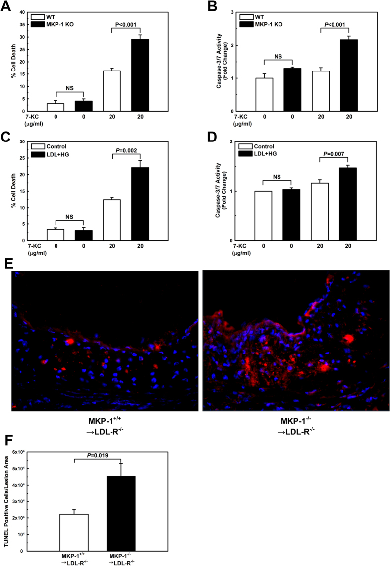
Figure 4. Both MKP-1-deficient and metabolically primed macrophages are sensitized to oxysterol-induced apoptosis.
Apoptosis was assessed in peritoneal macrophages treated with vehicle or 7-KC for 24 h. Cell death was measured by trypan blue dye exclusion and caspase 3/7 activation in peritoneal macrophages isolated from wildtype (WT) and MKP-1−/− (KO) mice (A+B), and in unprimed (Control) and metabolically primed (LDL + HG) peritoneal macrophages from C57/BL6 mice (C+D). Results are shown as mean ± SE (n = 3–4). Cell death in the atherosclerotic aortic roots of mice that received either wildtype or MKP-1−/− bone marrow and were fed a high-fat diet for 12 wk was assessed by TUNEL-positive cells relative to the atherosclerotic lesion area (E+F). Experiments were performed using 5–8 mice per experimental group (MKP-1+/+ → LDL-R−/−: n = 5; MKP-1−/− → LDL-R−/−: n = 8). Results are shown as mean ± SE.

