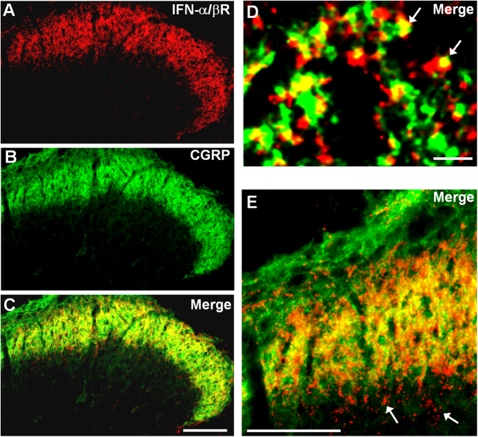
Figure 1. Expression of IFN-α receptors in the spinal cord dorsal horn.
(A–C) Double staining of Type I-IFN receptor (IFN-α/βR) and CGRP in the superficial dorsal horn. Scale, 100 μm. (D,E) High magnification images showing colocalization of IFN-α/βR and CGRP in primary afferent terminals in the superficial dorsal horn (laminae I-IIo). Arrows in (D) indicate double-labeled terminals. Arrows in E show IFN-α/βR labeling in inner lamina II (IIi). Scales, 3 μm (D) and 100 μm (E).

