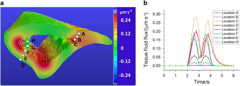
Figure 7.
Load-induced fluid flux at tissue level. (a) The distribution of the fluid flux magnitude and the flow direction at t=2.6 second during the loading phase. (b) The temporal changes of the fluid flux at several selected locations in both cortical (locations A–F) and trabecular sites (locations G–H).

