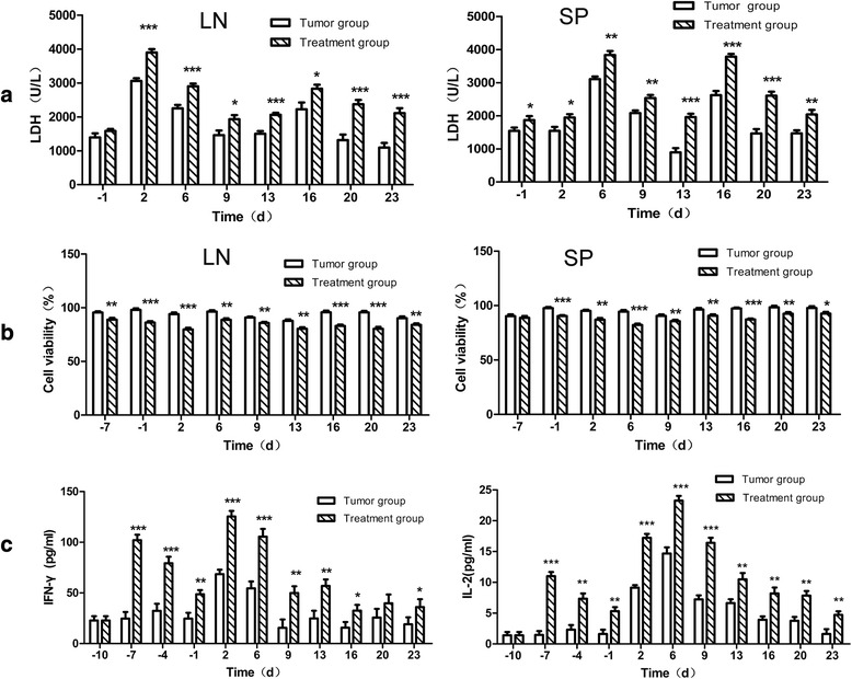
Fig. 4.

Tumor specific CTL activity in peripheral lymphoid organs and cytokines secretion in the serum The peripheral immune organs (LN and SP) were collected (3 mice/per group), and CD3+ T cells were isolated. These cells were incubated with CT26.WT tumor cells. Panel (a), represent the LDH release induced by T cells isolated from LN (left) and SP (right) of tumor control or treatment group mice. Panel (b), represents the CT26.WT tumor cell proliferation as measured by MTT assay post incubation with effector T cells at an E: T ration of 50:1. Panel (c), represents the secretion of cytokines measured by ELISA kit from the serum of tumor control and treatment group mice. Data are represented as mean ± SD. *denotes p < 0.05,**denotes p < 0.01, ***denotes p < 0.001
