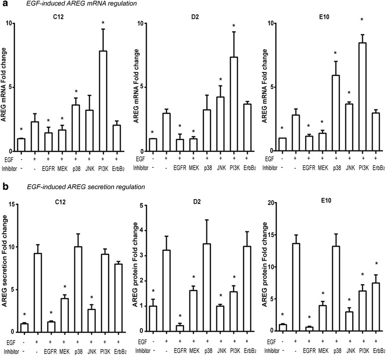
Fig. 5.

The intracellular pathways in EGF-induced AREG expression. The EGF-induced AREG mRNA (a) and protein (b) expression was reduced after EGFR-kinase inhibition in all cell lines, while it showed no reduction after ErbB2-kinase inhibition and was differently affected by the MAPK inhibitors. a Whereas MEK-inhibition reduced EGF-induced AREG mRNA expression in all the three cell lines, p38- and JNK-inhibition did not. PI3K-inhibition increased EGF-induced AREG expression in all cell lines. b EGF-induced AREG protein secretion could be totally blocked by EGFR- inhibition. Whereas MEK- and JNK-inhibition profoundly decrease EGF-induced AREG expression in all cell lines, p38-inhibition had no effect. The PI3K-inhibitor reduced EGF-induced AREG production in the conventional OSCC cell line D2 and E10, but had no effect on the basaloid OSSC cell line C12. The ErbB-2- inhibition inhibited the AREG increase in E10 cell line, only. *represents significant difference with EGF stimulation groups without inhibitors (student’s t-test)
