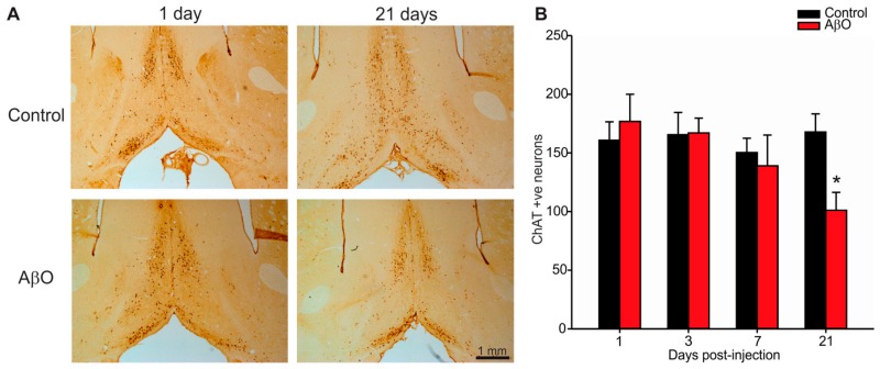
Figure 3.
Immunolabelling for cholinergic neurons within the basal forebrain. Paraformaldehyde perfused rat brains were sectioned at 30 µm and stained with the ChAT antibody that specifically labels cholinergic neurons within the basal forebrain. (A) Photomicrographs of the basal forebrain in coronal rat brain sections from AβO-injected and PBS-injected (control) rats 1, and 21 days post-injection. Scale bar is 1 mm; (B) Quantification of cholinergic neuron cell counts from three adjacent tissue sections per animal. AβO-injected rats had significantly more ChAT labelling in the basal forebrain compared to controls 21 days post-injection. Data presented as group means ± SEM. * indicates statistical significance between AβO-injected and control rats using 2-way ANOVA followed by Tukey’s post hoc analysis, p < 0.05, n = 5 for each experimental group.

