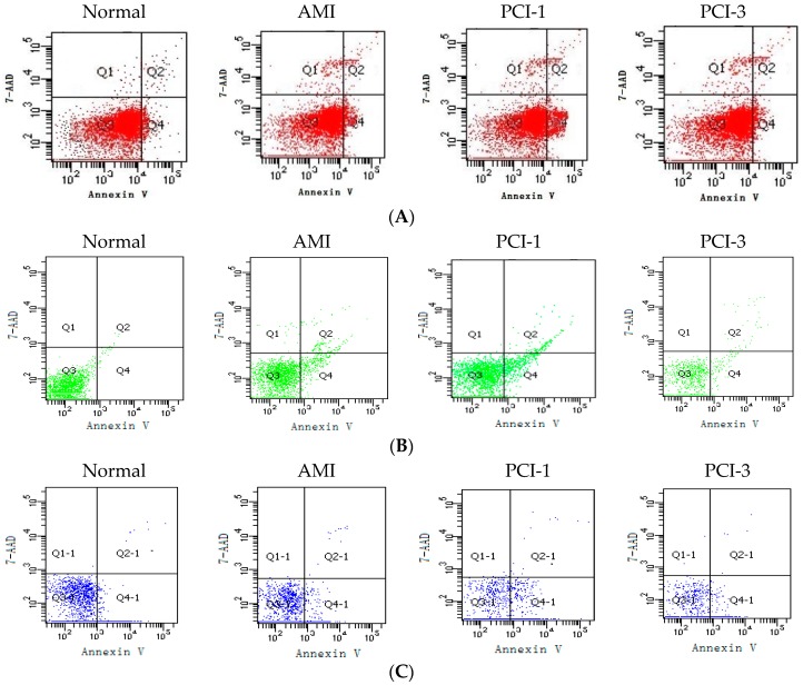
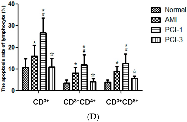
Figure 2.
Apoptosis rate of T lymphocytes. FACS results showed that the apoptosis rate of all the T cells of CD3+ labeled with APC (A); CD3+CD4+ labeled with PE (B); and CD3+CD8+ labeled with APC-Cy7 (C) increased at the onset of AMI and PCI-1, especially the CD3+ lymphocytes. However, they all returned to normal levels at PCI-3. The apoptosis rate of all these T cells was quantified by densitometry (n = 20) (D). * p < 0.05 vs. Normal group; # p < 0.05 vs. AMI group; ☆ p < 0.05 vs. PCI-1 group.


