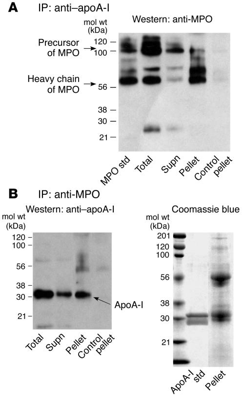
Figure 4.
Coimmunoprecipitation of MPO and apoA-I in plasma. (A) ApoA-I immunoprecipitation (IP) of plasma, followed by MPO Western blot (10–20% SDS-PAGE). (B) MPO IP of plasma, followed by apoA-I Western blot (10–20% SDS-PAGE) (left) or Coomassie blue staining (right). The MPO Western blot (A) was probed with rat anti–human MPO and includes, in the first lane, isolated human MPO as standard. The anti-MPO antibody used predominantly recognizes the heavy chain of MPO and thus highlights both heavy chain and precursor protein forms of MPO. The apoA-I Western blot (B) was probed with goat anti–human apoA-I. For each Western blot, the lanes were loaded with 10 μg of protein from plasma, the IP supernatant (Supn), and the specific and control immune complexes recovered from the IP pellets, in the indicated sequential order.

