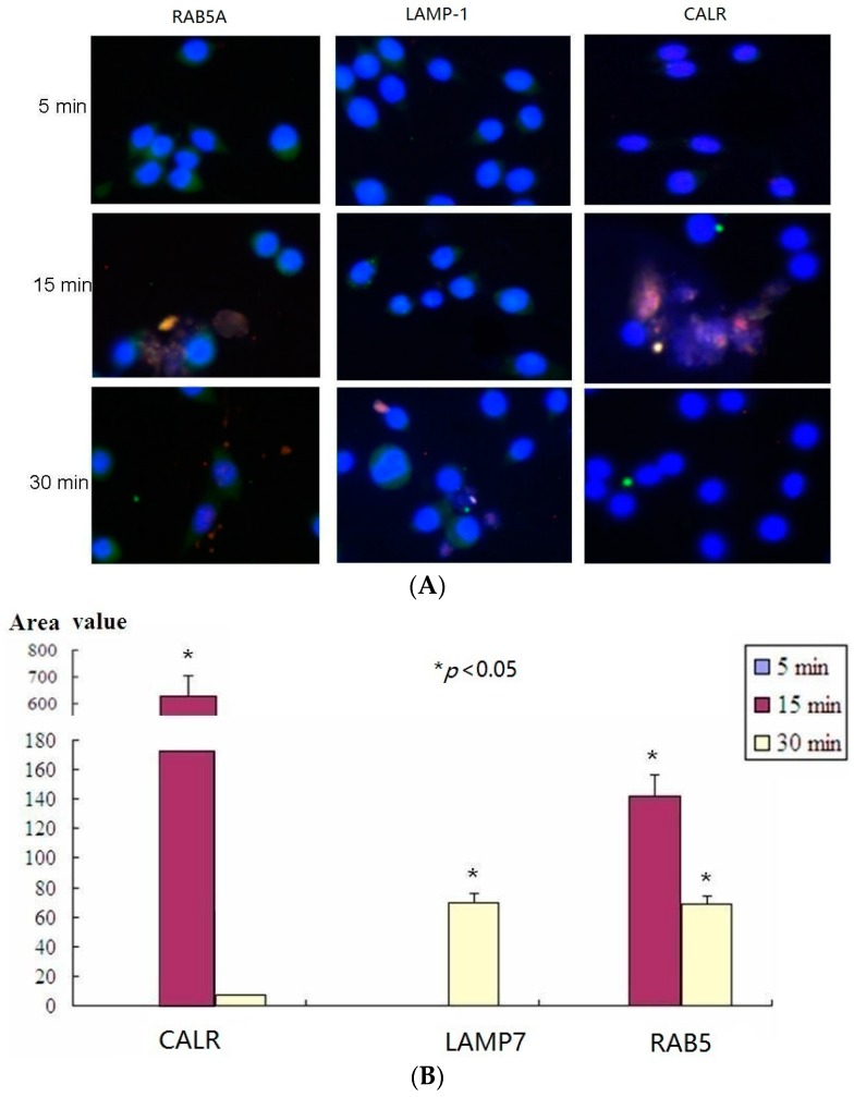
Figure 4.
Results of confocal microscopy assay. (A) mø264.7 cells were treated with LTB and fixed at 5, 15, or 30 min, respectively. After penetration of 0.5% Triton-X 100, the samples were blocked with 10% BSA/PBS and incubated with the rabbit-anti-RAB5A, rabbit-anti-LAMP-1, rabbit-anti-CALR, and mouse-anti-His-tag. Then the samples were incubated with secondary DyLight 488 labeled goat anti-rabbit IgG and Cy3 labeled goat anti-mouse IgG to detect the RAB5A, LAMP-1, CALR (green) and LTB (red), respectively. Images were taken using a confocal laser scanning microscope (FluoView1000, Olympus, Tokyo, Japan) with a 20× objective using the sequential scanning mode (200×) and processed using the FluoView software (Olympus); and (B) Quantitative analysis of microscopy images (* p < 0.05).

