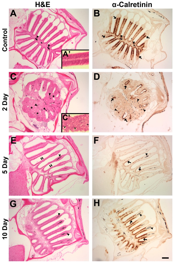
Figure 2.
The morphology of the olfactory organ and distribution of olfactory sensory neurons were examined with histological and immunohistochemical techniques. Unlesioned control olfactory organs sectioned in the horizontal plane displayed a semi-symmetrical shape with radiating lamellae (A) and typical olfactory epithelium appearance at higher magnification (A′); there was dense anti-calretinin labeling along the sensory epithelium ((B), arrows). Pigment cells were apparent in the lamina propria (arrowheads); (C) olfactory organs 2 days after intranasal infusion with 1 M zinc sulfate exhibited inflammation and fusion of lamellae (asterisks), and pigment was more dispersed (arrowheads). (C′) Higher magnification revealed vacuoles (v) and a generally disorganized epithelium; (D) anti-calretinin labeling (arrows) was diminished and confined to the apical surface of the epithelium. By 5 days after 1 M zinc sulfate exposure, the sensory epithelium was noticeably thinner than control tissue ((E), double arrows), and anti-calretinin labeling showed there were numerous olfactory sensory neurons dispersed throughout the tissue ((F), arrows). Pigment cells were again confined to the lamina propria (arrowheads). The morphology of the olfactory organ 10 days after 1 M zinc sulfate application more closely resembled that of control (G), and anti-calretinin labeling appeared similar to control levels in amount and intensity ((H), arrows). Scale bar = 100 μm (A–H) or 25 µm (A′,C′).

