Figure 4.
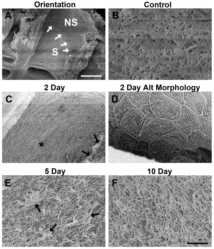
Scanning electron microscopy allowed analysis of the effects of zinc sulfate exposure on surface structures. (A) The sensory (S) and non-sensory (NS) regions of a lamella are distinguished with a defined separation (white arrows) in control fish; (B) the surface of control sensory epithelia was densely packed with cilia from OSNs, which obscured viewing of the shorter microvilli that are also present on the apical surface; (C) at 2 days after zinc sulfate exposure, the sensory epithelial surface appeared to contain only microvilli, with no evidence of cilia (*). Cilia in the non-sensory epithelium remained (black arrows); (D) an alternate morphology with no cilia or microvilli was seen at 2 days in 25% of the specimens examined. In these specimens, only microridges were apparent; (E) on the surface of the sensory epithelium of fish examined 5 days after infusion with the toxicant, intermittent cilia (black arrows) were present across the mat of microvilli; (F) by 10 days of recovery, the sensory epithelium appeared to be densely packed with cilia and microvilli, similar to control tissue. Scale bar in (A) = 100 μm; scale bar in (F) = 7 μm (B–F).

