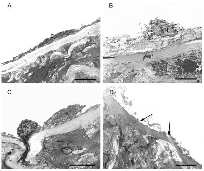
Figure 3.
The figure shows the endothelium in the CON, SCH, and CH groups. (A) Normal endothelial cell (E) in the CON group; (B) Dissolved endothelial cell membrane and abnormal nuclear (N) feature in the SCH group; (C) Dissolved endothelial cell membrane and heterogeneous chromatin edge accumulation in the CH group; (D) Endothelial cells shed and parts of the elastic membrane exposed in the CH group (→). Scale bar = 4 µm.

